Introduction
The Triangular Fibrocartilage Complex (TFCC) plays a critical role in wrist stability and function, yet injuries to this vital structure can lead to significant pain, reduced mobility, and limitations in daily activities. If left untreated, a TFCC injury can cause chronic discomfort and long-term disability. This blog provides a comprehensive guide to understanding TFCC injuries, their causes, diagnosis, and advanced treatment options, including cutting-edge regenerative therapies.
What Is the TFCC?

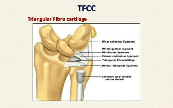
Anatomy of the TFCC
The TFCC is a complex structure located on the ulnar side of the wrist (pinky finger side). It consists of:
- Fibrocartilage Disc (Articular Disc): Acts as a cushion between the ulna and the carpal bones.
- Ligaments: Including the dorsal and palmar radioulnar ligaments, which stabilize the distal radioulnar joint (DRUJ).
- Sheath of the Extensor Carpi Ulnaris (ECU): Provides additional wrist stabilization.
- Meniscus Homologue: A soft tissue structure aiding in shock absorption.
Functions
- Stabilizes the DRUJ.
- Distributes axial load across the wrist.
- Enables smooth wrist rotation (pronation and supination).
Causes of TFCC Injuries
TFCC injuries can occur due to
Trauma
- A fall on an outstretched hand (FOOSH injury).
- Direct impact to the wrist during sports or accidents.
Repetitive Strain
Activities requiring repetitive wrist motions (e.g., racket sports, manual labor).
Degeneration (Wear and Tear)
- Common in older adults due to natural wear of the fibrocartilage.
- Linked with conditions like ulnar impaction syndrome, where the ulna is abnormally long, causing excessive pressure on the TFCC.
Rheumatoid Arthritis
Inflammatory conditions can weaken the TFCC, making it prone to injury.
Grades of TFCC Tears

Palmer classification of TFCC lesions
TFCC injuries are categorized based on the Palmer Classification System
Type 1: Traumatic Tears
- 1A: Tear of the central disc.
- 1B: Avulsion (tearing away) of the ulnar side of the TFCC.
- 1C: Disruption of the ulnar carpal ligaments.
- 1D: Avulsion of the radioulnar ligament from the radius.
Type 2: Degenerative Tears
- 2A: TFCC degeneration without perforation.
- 2B: TFCC degeneration with thinning.
- 2C: TFCC thinning with perforation.
- 2D: TFCC thinning with perforation and chondromalacia (cartilage damage) of the lunate.
- 2E: Advanced degeneration involving the ulnar head and DRUJ instability.
Symptoms of TFCC Injury
- Pain: On the ulnar side of the wrist, worsened by twisting motions (e.g., opening jars, turning keys).
- Swelling: Localized to the wrist.
- Clicking or Popping Sensation: During wrist movement.
- Grip Weakness: Difficulty gripping objects due to pain or instability.
- Instability: Feeling of looseness in the wrist joint.
- Reduced Range of Motion: Especially during pronation (palm-down motion) or supination (palm-up motion).
Diagnosis of TFCC Injury
Clinical Examination
- Fovea Sign: Tenderness over the ulnar wrist near the DRUJ.
- Stress Tests: Pain reproduced during ulnar deviation or rotational movements.
- Grip Strength Testing: Reduced strength compared to the unaffected wrist.
Imaging Techniques
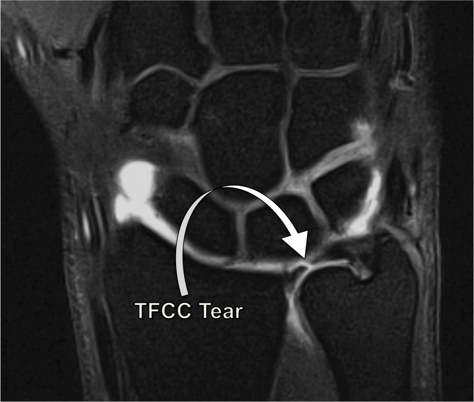
MRI (Magnetic Resonance Imaging)
- Considered the gold standard for diagnosing TFCC injuries.
- Provides high-resolution images of soft tissue structures, including tears, degeneration, and joint instability.
- Can identify associated conditions like ulnar impaction syndrome.

MRI of the wrist showing a TFCC tear
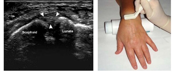
Diagnostic Ultrasound
Useful for dynamic assessment of TFCC structures. Can detect ligament tears, fluid accumulation, and ECU sheath abnormalities in real-time.

Diagnostic Ultrasound showing the TFCC ligament
X-Ray
- Typically normal but used to assess bone alignment and ulnar variance.
- Helps rule out fractures or arthritis.
Arthroscopy
A minimally invasive surgical procedure to directly visualize and confirm TFCC injuries when imaging is inconclusive.
Treatment Options for TFCC Injuries
Conservative Management
- Rest and Immobilization: Wrist brace or splint to limit movement and reduce strain.
- Non-Steroidal Anti-Inflammatory Drugs (NSAIDs): Reduce pain and inflammation.
- Physical Therapy: Exercises to strengthen wrist muscles and improve range of motion, Proprioceptive training to restore joint stability.
Regenerative Treatments
Regenerative therapies aim to repair damaged tissues and restore function without invasive surgery. Generally indicated for mild to moderate grades of tears.
Platelet-Rich Plasma (PRP) Therapy PRP, derived from the patient’s own blood, contains concentrated growth factors that promote healing and tissue repair.Ultrasound-guided injection into the TFCC to ensure precise delivery.
Benefits
- Reduces inflammation.
- Accelerates the healing process.
- Effective for partial TFCC tears and early degeneration.
Prolotherapy
Involves injecting a dextrose solution to create a controlled inflammatory response, stimulating tissue regeneration and strengthening ligaments.Ultrasound guidance ensures accurate delivery to the injured TFCC.
Benefits
Improves ligament stability and wrist function.
Surgical Intervention
Surgery is indicated for severe TFCC tears, chronic instability, or when conservative and regenerative treatments fail.
- Arthroscopic Debridement: Removes damaged tissue to reduce pain and improve joint mechanics.
- Arthroscopic Repair: Sutures are used to repair torn ligaments or disc structures.
- Ulnar Shortening Osteotomy: Indicated for ulnar impaction syndrome, this procedure shortens the ulna to relieve pressure on the TFCC.
- DRUJ Stabilization: Used to restore stability in cases of DRUJ instability associated with TFCC injury.
When to Seek Medical Help
If you experience persistent wrist pain, swelling, or reduced mobility despite home treatments, consult a specialist. Early diagnosis and intervention can prevent complications and ensure better outcomes.
Conclusion
TFCC injuries can significantly affect wrist function, but early diagnosis and appropriate treatment can lead to full recovery. At Alleviate Pain Clinic, we offer state-of-the-art diagnostic tools and advanced regenerative therapies to help you regain wrist strength and mobility. Contact us today to schedule a consultation.
References
- Palmer AK, Werner FW. The triangular fibrocartilage complex of the wrist—anatomy and function. J Hand Surg Am. 1981.
- Bednar MS, Arnoczky SP, Weiland AJ. The microvasculature of the triangular fibrocartilage complex: Its clinical significance. J Hand Surg Am. 1991.
- Sagerman SD, Short WH. The role of MRI in the diagnosis of TFCC injuries. Radiographics. 1998.
- Horton T, Wojtys EM. Arthroscopic management of TFCC injuries. Clin Sports Med. 1995.
- Ahmed I, Salmon J. Advances in regenerative medicine for wrist injuries: PRP and stem cell therapies. Orthop Res Rev. 2022.
- Shinohara T, Nakamura R. Diagnostic ultrasound in the evaluation of wrist injuries. Ultrasound Med Biol. 2010.
- Adams BD. Partial excision and repair of TFCC tears: Indications and techniques. Hand Clin. 2012.
- Dutton C, Hoskins WT. Prolotherapy for wrist instability: Clinical outcomes. J Orthop Sci. 2019.
- Thomas BP, Sreekanth R. Role of PRP in managing TFCC degeneration. World J Orthop. 2017.
- Matsuki K, Doi K. Arthroscopic versus open repair for TFCC tears: A review. J Hand Surg Eur. 2018.
FAQs About TFCC injury
The TFCC is a triangular structure of cartilage and ligaments in the wrist that stabilizes the ulnar side of the wrist and supports rotation and load distribution.
TFCC injuries are typically caused by trauma (e.g., falls on an outstretched hand), repetitive wrist motions, or degenerative wear and tear.
Common symptoms include pain on the pinky side of the wrist, swelling, clicking or popping sounds during movement, and reduced wrist strength or stability.
TFCC injuries are relatively common in athletes, individuals performing repetitive wrist movements, and older adults experiencing degenerative changes.
A combination of clinical examinations, imaging techniques like MRI or ultrasound, and patient history is used to diagnose TFCC injuries.
MRI provides high-resolution images of soft tissues, helping to detect even minor tears or degeneration in the TFCC.
While X-rays can’t visualize soft tissues, they help rule out fractures, arthritis, or bone-related abnormalities that might contribute to wrist pain.
Ultrasound is useful for real-time dynamic imaging, assessing ligament tears, fluid buildup, and associated structures.
Mild or partial TFCC tears often heal with conservative treatment, including rest, splinting, physical therapy, and NSAIDs.
A wrist brace that immobilizes the ulnar side of the wrist and limits rotational movement is often recommended.
Recovery times vary but generally range from 6 to 12 weeks for mild injuries.
Proprioceptive exercises, grip-strengthening routines, and gentle range-of-motion exercises prescribed by a physical therapist are beneficial.
Platelet-Rich Plasma (PRP) therapy involves injecting a concentration of platelets derived from your blood into the injured TFCC to stimulate healing.
Prolotherapy uses dextrose injections to trigger a mild inflammatory response, encouraging tissue regeneration and strengthening ligaments.
Studies suggest that regenerative treatments can be highly effective for partial tears and degenerative TFCC injuries, especially when guided by ultrasound.
Ultrasound guidance ensures precise delivery of injections to the injured area, improving effectiveness and reducing risks.
Surgery is indicated for severe tears, DRUJ instability, or injuries unresponsive to conservative and regenerative treatments.
Options include arthroscopic debridement (removal of damaged tissue), arthroscopic repair (suturing torn ligaments), and ulnar shortening osteotomy for ulnar impaction syndrome.
Recovery typically takes 3 to 6 months, depending on the procedure and the severity of the injury.
Risks include infection, stiffness, and incomplete healing, though these are minimized with proper surgical techniques and post-operative care.


