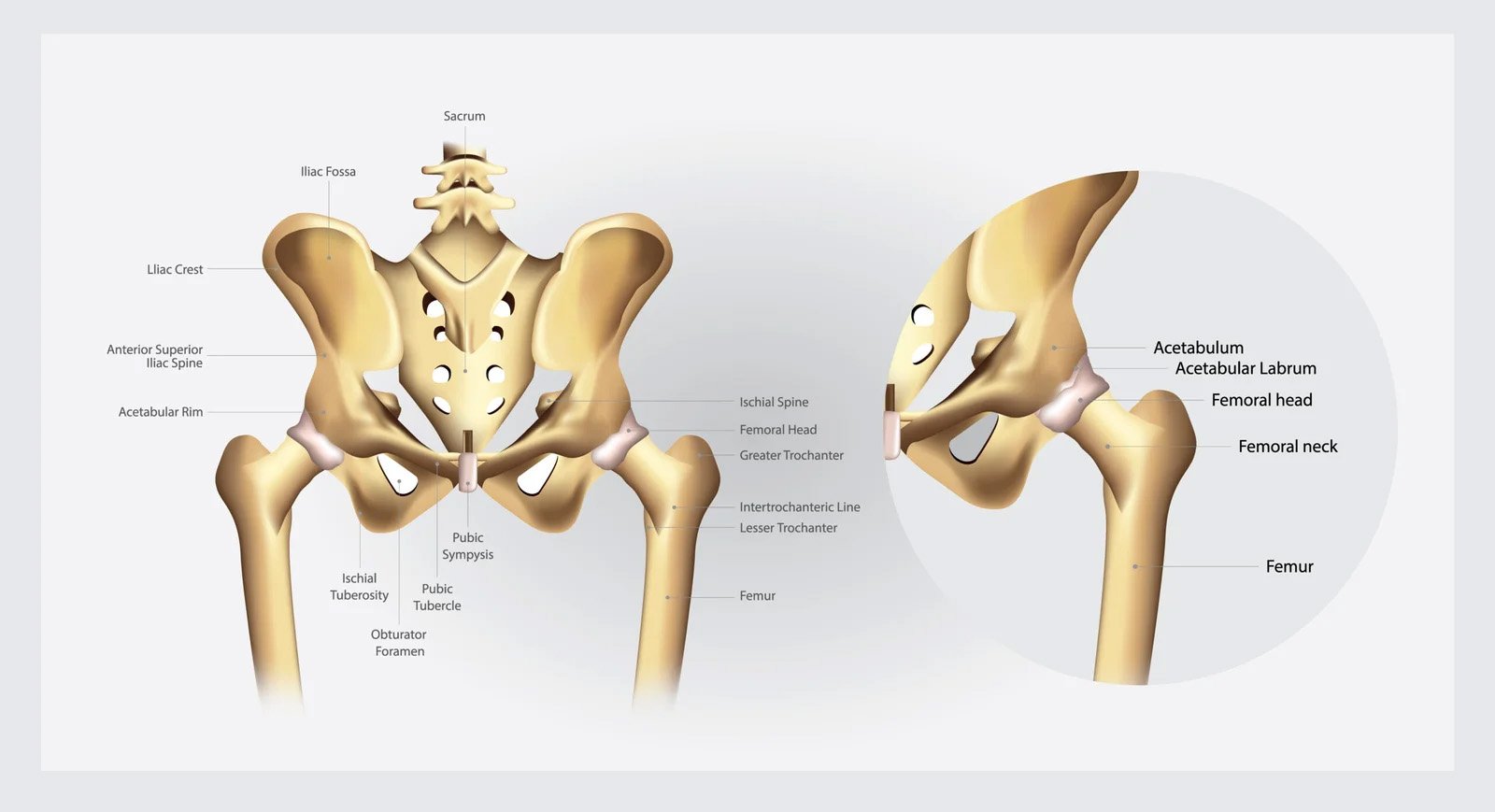
Understanding Hip Anatomy
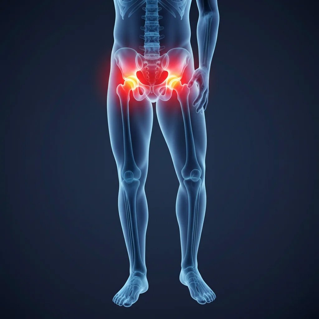
The hip is a ball and socket joint which is present between the head of the femur and the acetabulum of the pelvis. Cartilage and a lubricating membrane known as the synovial membrane cushion motion. Movement is stabilized by surrounding muscles (gluteals, hip flexors), ligaments (iliofemoral, pubofemoral), tendons, and bursae. Sensation is provided by nerves such as the sciatic and femoral. This anatomy is crucial to understand, as no matter what the problem is: hip arthritis, hip bursitis, hip tendonitis, or hip pain, a precise program is crucial to treat the hip pain without surgery.
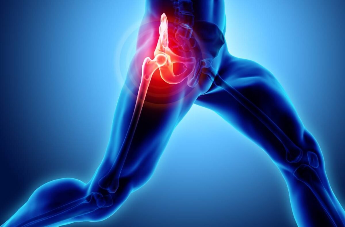
Best Clinic in Bangalore for Hip Pain Conditions
Alleviate Pain Clinic presents a destination centre for hip condition treatment in Bangalore. Our purpose is to offer high-tech diagnostics and precise treatment with a non-surgical treatment approach for hip pain. The treated conditions are:
When to See a Doctor for Hip Pain
Schedule an appointment with a specialist in case the hip pain:

Lasts longer than two weeks.

Gets worse at night or restricts walking.

Increased stiffness, swelling, or tenderness.

Radiates to the groin or the thigh region.

It is accompanied by fever or weight loss.
Diagnosis of Hip Pain
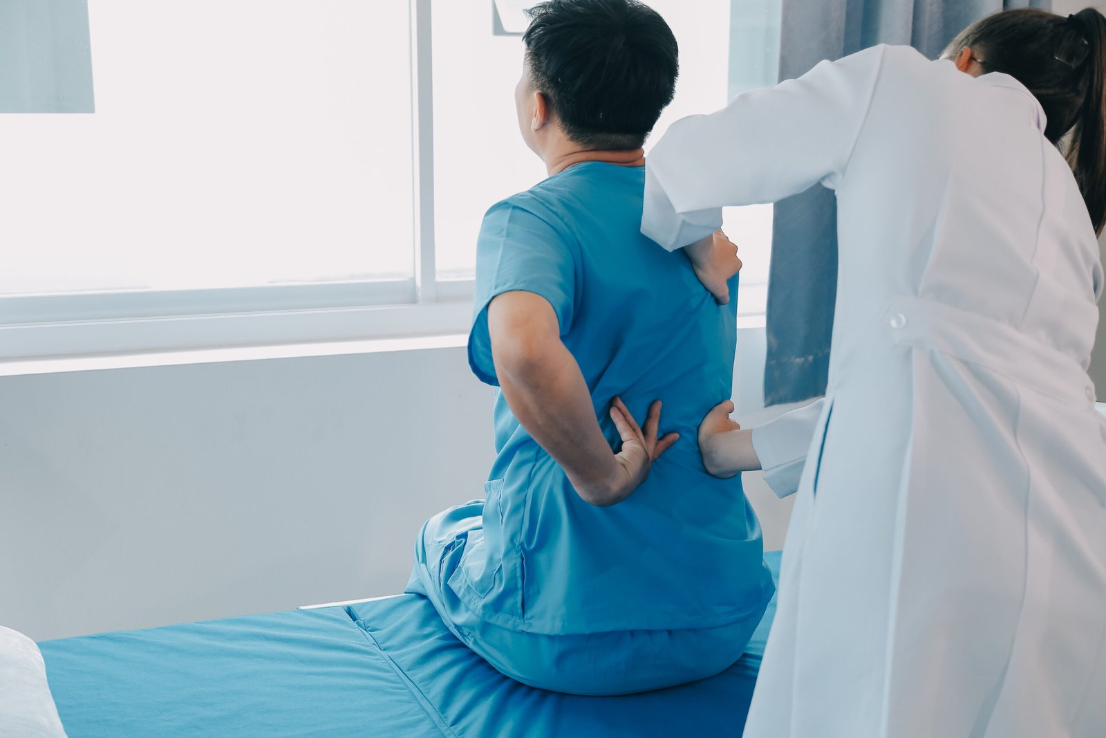
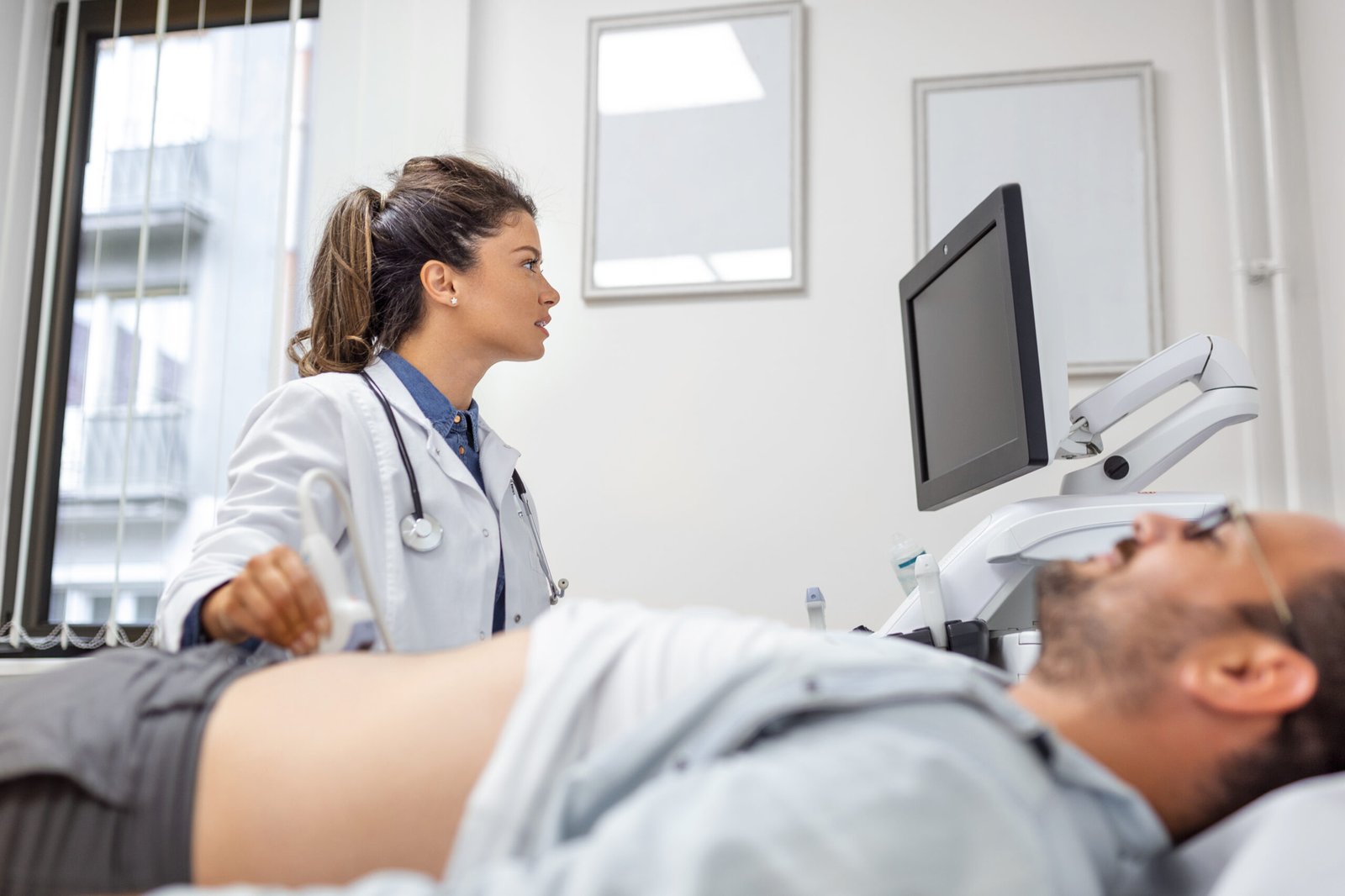
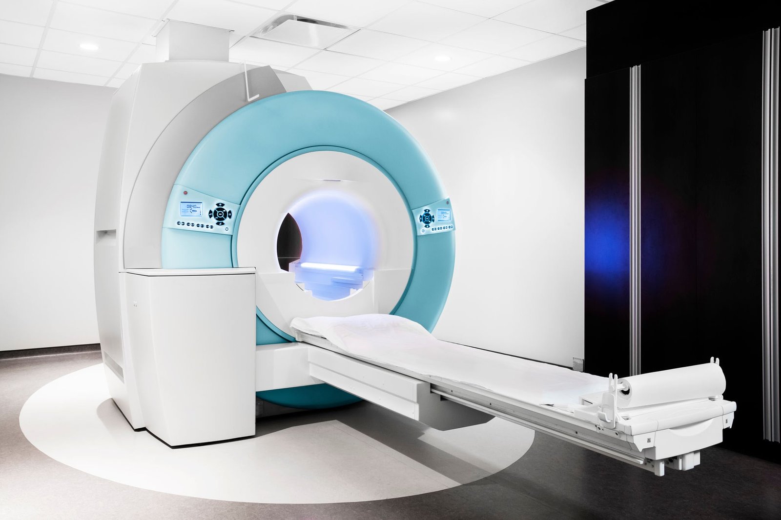
A precise diagnosis of the hip pain is a crucial factor in hip pain management. At Alleviate Pain Clinic, we believe in treating hip pain without surgery and address these issues by using a combination of clinical assessment, imaging techniques, and movement analysis, which allows our specialists to identify the root cause of the pain- joint degeneration, bursitis, soft tissue damage, or nerve-related pain.
Our Approach To Non-Surgical Hip Pain Treatment
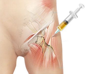
Nerve Blocks
Nerve blocks involve precisely injecting an anesthetic near specific nerves to interrupt pain signals. This procedure provides immediate relief and helps us diagnose the exact source of your discomfort.
PRP (Platelet Rich
Plasma Therapy)
PRP therapy uses a concentration of your own blood platelets to accelerate the healing of injured tendons, ligaments, muscles, and joints, promoting natural tissue repair and reducing inflammation.
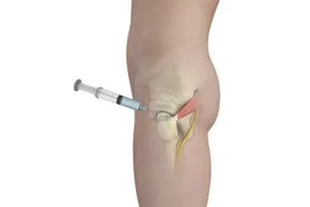
Prolotherapy
Prolotherapy is a regenerative injection therapy that stimulates the body’s natural healing process to strengthen and repair weak or injured ligaments and tendons, providing long-term pain relief.
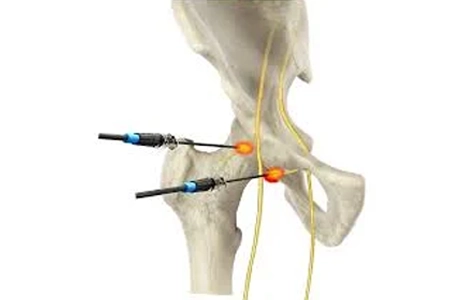
Radiofrequency
This minimally invasive procedure uses heat generated from radio waves to target specific nerves, effectively blocking pain signals from reaching the brain and offering lasting relief from chronic conditions.
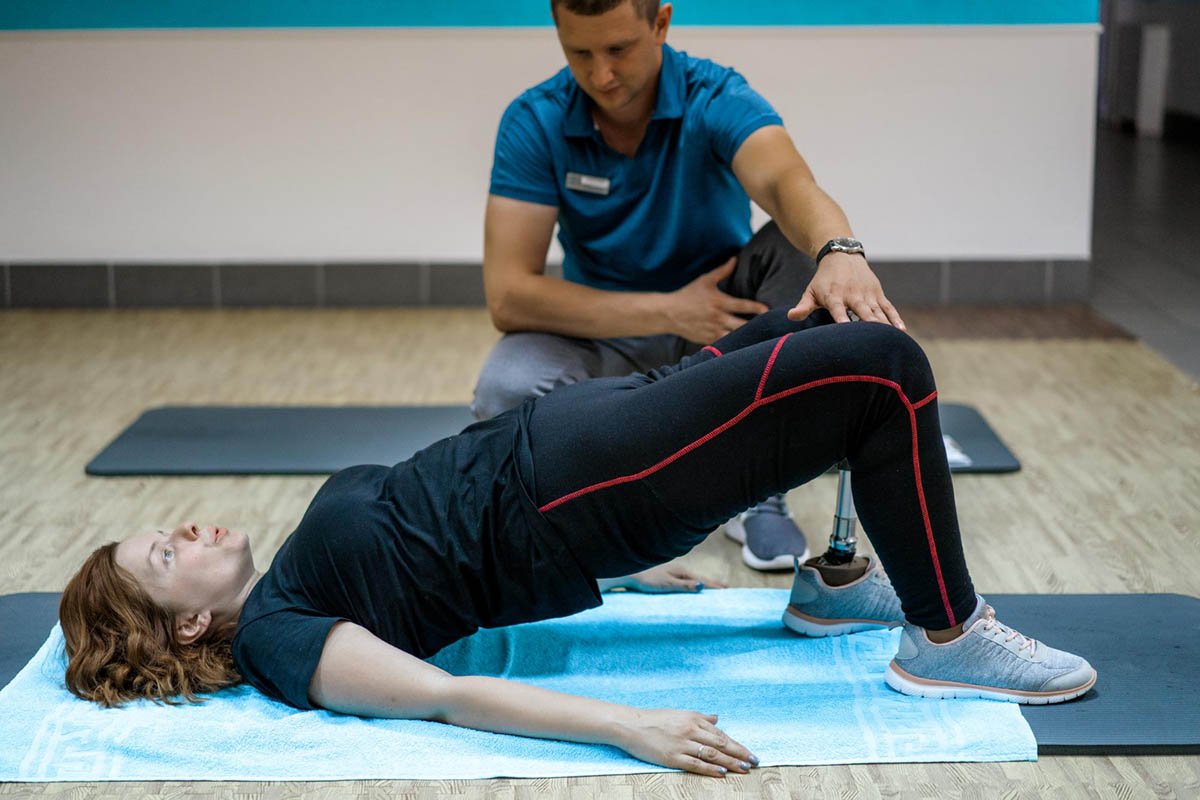
Physiotherapy
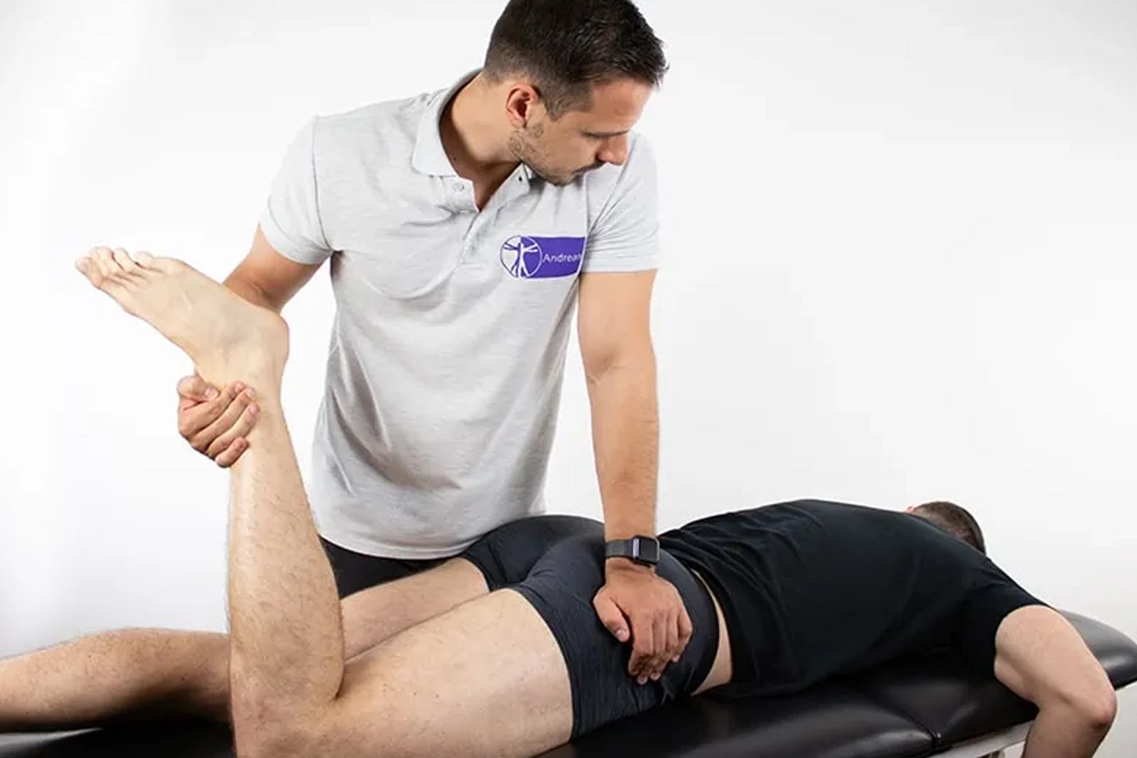
Our customized physiotherapy programs use targeted exercises and manual therapy to restore movement, improve strength, and reduce pain, empowering you to regain function and prevent future injuries.

Lifestyle Changes
We guide you in making sustainable lifestyle adjustments, including nutrition, exercise, and stress management, to support your treatment, enhance recovery, and improve your overall quality of life.
How Are We Different from Other Clinics and Orthopedics?

Experienced Doctors
At Alleviate Pain Clinic, we also have the best hip pain specialists in Bangalore. The medical professionals are widely educated on the diagnosis and treatment of numerous hip disorders. Patients can get the correct diagnosis and get effective individual solutions to their problems due to the specialisation in musculoskeletal medicine and interventional pain care.

Personalised & Advance Treatments
We think each hip condition is different. And that is why we provide individually tailored care plans based on the most innovative and non-invasive treatments such as PRP, stem cell therapy, and regenerative medicine. Such advanced treatments are tailor-made to patients and are less invasive since they involve no surgical operations, but help fight inflammation and restore normal joint movement and functionality with minimum downtime.

Non-Surgical Treatments
Alleviate Pain Clinic believes in curing hip-type pain with non-surgical procedures as a priority to make sure the process is quick and less risky. Among our treatment options, we have needled injections, physiotherapy, prolotherapy, and regenerative medicine. The reduction of pain levels, increased mobility, and getting a long-lasting relief without replacement of the hip and invasive surgery make the patients benefit from the procedure.
Our Doctors
Reviews
Our Clinics Gallery
Frequently Asked Questions
Arthritis, bursitis, tendinitis, and hip impingement, or overuse of muscles due to poor posture, are common causes of hip pain without injury. These disorders take time to develop and consequently lead to chronic discomfort that becomes worse with motion or sedentariness.


